May 14, 2026
WHEN WATER IS THE TOPIC, THE BUILDING IS PART OF THE SCIENCE
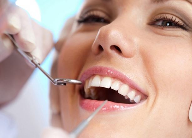
People usually arrive at a fluoride-in-water discussion through public health: dental outcomes, dosage, equity, and long-term exposure. What gets less attention is the moment water stops behaving like “tap water” and starts behaving like a building problem. A burst pipe, a flooded basement, a roof leak that runs behind the walls. In those moments, the questions change fast: what was in that water, what did it touch, what’s now airborne, and how do we make the space safe again?
That overlap is why we talked to the owner of Boston Water Damage Restoration while putting this piece together. Their team sees the real-world edge cases that rarely show up in abstract debates: families displaced by a leak, schools dealing with soaked materials, and homeowners trying to decide what needs professional remediation versus what can be handled with careful cleanup.
This article bridges fluoride-in-water concerns with a practical reality: the quality and safety of water is not only about treatment levels. It is also about what happens when water leaves the pipe, saturates a structure, and changes the indoor environment.
1) FLUORIDE CONVERSATIONS START WITH CONCENTRATION, NOT CONFUSION

Fluoride discussions can get emotionally charged because they sit at the intersection of health, trust, and policy. A helpful way to keep the conversation grounded is to separate three ideas: the recommended target level for community water systems, higher levels that may increase the likelihood of dental fluorosis during tooth development, and much higher levels that trigger regulatory concern.
In the United States, the U.S. Public Health Service recommendation commonly referenced by public health sources is 0.7 mg/L as a level intended to maximize cavity prevention benefits while minimizing the risk of dental fluorosis. The CDC’s overview of community water fluoridation recommendations summarizes that guidance clearly here: https://www.cdc.gov/fluoridation/about/community-water-fluoridation-recommendations.html.
That baseline matters because it frames the rest of the discussion. Most people are not interacting with “unknown chemistry.” They are interacting with a system that targets a specific concentration, measured and monitored, with ongoing debate focused on policy choices, cumulative sources, and individual risk tolerance.
2) WHEN WATER BECOMES “WATER DAMAGE,” THE RISK PROFILE CHANGES
Now shift the scenario. The issue is no longer the fluoride level in a glass of water. The issue is water where it does not belong: behind drywall, under flooring, inside insulation, or pooled in a basement. That changes what you need to think about, because the primary health risks often become microbial growth, particulate contamination, and long-term moisture problems that alter indoor air.
In other words, the biggest health question after a leak is usually not “what was the fluoride level.” It’s “what is going to grow here, what is going to linger, and what is going to get into the air we breathe.”
The owner we spoke with described it simply: clean water does not stay clean once it moves through building cavities and porous materials. It picks up debris, contacts dust, and leaves behind moisture in spaces that were never designed to dry quickly. This is where prompt, methodical restoration matters, because time is the enemy of indoor health after a water event.
3) THE “HIDDEN” HEALTH EFFECTS OF MOISTURE: WHY DRYING IS A HEALTH DECISION
A surprisingly common misconception is that if something looks dry, it is dry. Building materials can hold moisture deep inside, and that moisture can persist long enough to create conditions for mold growth or other indoor air quality issues.
The EPA’s guidance on mold and moisture emphasizes the practical core of prevention: control moisture, dry promptly, and fix the underlying water problem so the issue does not repeat. This guide is a strong reference point for why drying and remediation are part of health protection, not just cosmetic repair: https://www.epa.gov/mold/brief-guide-mold-moisture-and-your-home.
For readers of a fluoride-focused publication, this matters because it expands the water conversation. Water affects health through chemistry, yes. But it also affects health through physics and biology when it changes the indoor environment.
4) WHAT RESTORATION PROFESSIONALS ACTUALLY DO AFTER A WATER EVENT
A good restoration workflow is not simply “remove the wet stuff.” It’s a sequence designed to prevent secondary harm: trapped moisture, persistent odor, recurring microbial growth, and compromised materials that keep shedding particles.
From the perspective of Boston Water Damage Restoration, the most important steps are often the least visible: moisture mapping, controlled drying, and verification. That is especially true in older buildings where water can travel along framing, wick into plaster, or settle into low points that never fully dry without intervention.
This is also where trust and documentation matter. If a family has concerns about water quality or chemical exposure, a clear remediation record helps them understand what happened, what was removed, what was dried, and what was tested for moisture before the space was returned to normal use.
5) BRIDGING THE “WATER QUALITY” MINDSET WITH THE “BUILDING HEALTH” MINDSET
Fluoride discussions often revolve around long-term, low-dose exposure. Water damage conversations are usually short-term, high-disruption events. But the bridge between them is the same core principle: dose and duration matter, and context changes risk.
A few examples make this easier to see:
- A stable municipal water supply can be managed through monitoring and public reporting.
- A water-damage event is chaotic, and the variables multiply quickly: source, category, contact surfaces, time-to-dry, and whether materials were removed or sealed.
- After a major leak, the bigger health lever is often indoor air management and moisture control, because that affects daily exposure immediately.
This is not a reason to dismiss fluoride questions. It’s a reason to recognize that “water and health” is bigger than a single parameter.
6) A PRACTICAL CHECKLIST FOR READERS WHO WANT ACTIONABLE GUIDANCE
If you want a health-first approach that respects both water quality and indoor environment risk, these steps are a strong starting point after any significant water event:
- Identify the source and stop it, then document what happened and when.
- Assume porous materials may hold moisture longer than they appear to.
- Prioritize fast drying and dehumidification, especially in wall cavities and under flooring.
- Remove materials that cannot be reliably dried or that have been contaminated.
- Fix the underlying cause, including drainage, plumbing, or envelope issues, to prevent recurrence.
- Use reputable restoration professionals when the affected area is large, hidden, or time-sensitive.
This is the kind of practical, prevention-oriented work that restoration teams do daily, and it aligns with the broader public health principle behind water discussions: reduce avoidable exposure, and reduce avoidable uncertainty.
7) THE TAKEAWAY: WATER SAFETY IS A SYSTEM, NOT A SINGLE NUMBER
Fluoride debates often focus on a number in a report. Water damage forces us to see the full system: infrastructure, building materials, indoor air, and human behavior under stress. Both perspectives matter, and both benefit from calm, evidence-based thinking.
If we want the healthiest outcome for communities, we should treat water as more than a treatment topic. We should treat it as a lifecycle topic, from distribution to the moment it interacts with our living spaces. That is why conversations about fluoride-in-water and conversations about restoration after leaks can sit in the same article without feeling forced. They are both about preventing harm, using good information, and responding appropriately when conditions change.
August 7, 2025
From Eyes to Teeth: The Ultimate Facial Aesthetic Makeover in Dallas, Texas
In the heart of Dallas, Texas, the trend of full facial aesthetic makeovers is gaining significant momentum. More individuals are seeking comprehensive transformations that address multiple areas of the face, not just isolated concerns. Among the most sought-after combinations are cosmetic dental treatments and blepharoplasty in Dallas (eyelid surgery). When performed in tandem or as part of a coordinated aesthetic plan, these procedures can rejuvenate the entire facial appearance, creating a more youthful, vibrant, and confident look.

Why Combine Blepharoplasty and Cosmetic Dentistry?
The face is a unified aesthetic canvas. While many people focus on individual features, true facial harmony comes from the balance between the eyes, smile, skin, and facial contours. This is why combining blepharoplasty and cosmetic dentistry can have such a powerful impact.
Blepharoplasty, commonly referred to as eyelid surgery, is a procedure that removes excess skin, fat, and muscle from the upper or lower eyelids. It addresses sagging eyelids, puffiness, and under-eye bags—issues that can make individuals appear tired, sad, or older than they are.
On the other hand, cosmetic dental procedures like veneers, crowns, whitening, and Invisalign can dramatically enhance the smile. A straight, white, and symmetrical smile has long been associated with health and attractiveness. When these procedures are done in isolation, they certainly improve specific areas. But when combined, they can completely transform how a person is perceived.
The Power of First Impressions
Studies consistently show that the eyes and the mouth are the first features people notice during an interaction. The eyes convey emotion and energy, while the smile reflects warmth and confidence. By enhancing both, patients can project a youthful, approachable, and self-assured image.
For Dallas professionals, public speakers, media personalities, and individuals in client-facing industries, the impact of these enhancements is not just personal but professional. Looking well-rested and confident can influence how others perceive competence, reliability, and vitality.
A Customized Approach for Every Patient
In Dallas, the approach to facial aesthetics is becoming increasingly personalized. Reputable clinics and practitioners are collaborating across disciplines—plastic surgery and cosmetic dentistry—to create tailored treatment plans that consider the whole face, not just one feature.
A patient may begin with a consultation at a dental clinic to discuss whitening and alignment, then be referred to a plastic surgeon to evaluate eyelid concerns. Alternatively, a patient seeking eyelid rejuvenation may be advised to consider a dental makeover to ensure the entire facial appearance is balanced and harmonious. The result is a more natural, refreshed look, rather than an obviously altered one.
Timing and Coordination Matter
While these procedures do not necessarily need to be performed at the same time, careful planning is essential. Some patients choose to undergo their dental treatments first, especially if orthodontics or bite alignment is involved, as this can change the way the lower face is structured. Once dental goals are achieved, blepharoplasty can be performed to fine-tune the overall aesthetic.
Others opt for blepharoplasty first, especially if drooping eyelids are interfering with vision or causing discomfort. After recovery, cosmetic dental treatments can enhance the lower part of the face, completing the transformation.
In either case, coordination between practitioners is key. Many Dallas clinics are now offering aesthetic concierge services, where a team of professionals work together to streamline appointments, recovery times, and aesthetic goals.
What to Expect: Recovery and Results
Blepharoplasty typically involves a few days of downtime, with swelling and bruising peaking around day two or three. Most patients return to normal activities within 7 to 10 days. Results are long-lasting and can dramatically improve the appearance of tired or aging eyes.
Cosmetic dental treatments vary widely in terms of recovery. Procedures like teeth whitening or bonding require no downtime, while veneers or orthodontics may involve several appointments over a few weeks or months. The good news is that dental treatments are usually non-invasive, so they can often be performed during or shortly after the recovery period from eyelid surgery.
When both procedures are completed, the results can be striking. A rejuvenated eye area paired with a bright, confident smile can take years off a person’s appearance and boost self-esteem.
Choosing the Right Professionals in Dallas
Dallas is home to a wide range of highly qualified plastic surgeons and cosmetic dentists. When considering a combined facial makeover, it is important to choose providers who not only specialize in their respective fields but are also open to interdisciplinary collaboration.
Look for surgeons who are board-certified and have extensive experience in facial aesthetics. Likewise, choose dentists who focus on cosmetic procedures and have a portfolio of before-and-after cases. Patient reviews, consultations, and open communication are critical to ensuring your aesthetic goals are understood and achievable.
Final Thoughts
In today’s image-conscious world, facial aesthetics are about more than vanity—they are about confidence, self-expression, and feeling good in your own skin. For Dallas residents seeking a complete facial transformation, combining blepharoplasty with cosmetic dental treatments offers a powerful and effective path toward renewal.
The journey from eyes to teeth isn’t just about looking better—it’s about becoming the best version of yourself. And with the expertise available in Dallas, the ultimate facial makeover is more accessible than ever.
November 4, 2023
Braces and Beams: What Baltimore Deck Repair Can Teach Us About Dental Alignment
In the charming city of Baltimore, where historic neighborhoods nestle against the bustle of modern life, the care we give to our homes—especially to features like outdoor decks—and the attention we pay to our health, particularly dental alignment, share a surprising parallel. This blog post delves into the fascinating similarities between the Baltimore deck repair service and the meticulous process of dental braces, drawing lessons from the former to shed light on the latter.

The Importance of a Strong Foundation
Deck Foundations and Dental Foundations
Before any repair work can commence on a Baltimore deck, one must ensure that the foundation is solid and stable. Similarly, before orthodontic braces are placed, a dentist must assess the health of a patient’s teeth and gums. The foundation for both a deck and a dental brace must be prepared to support the structure and adjustments that will follow.
Preventative Measures
In deck repair, preventative measures like sealing and staining can protect the wood from the harsh Chesapeake Bay weather. In dental care, fluoride treatments and sealants act as preventative care, protecting teeth from decay that could later complicate orthodontic treatments.
Aligning Structures
Straightening Beams and Teeth
When a deck’s beams become misaligned due to weather or wear, a skilled craftsman can realign and secure them to restore the deck’s integrity. Orthodontists use braces to apply steady pressure on teeth, guiding them into a proper alignment over time. Both processes require a period of adjustment, careful planning, and a commitment to maintenance.
The Role of Time
The alignment of deck beams doesn’t happen overnight; it requires time for the wood to settle and adapt to its new position. Braces, too, are not a quick fix. They move teeth gradually, and the process can take several months to a few years, depending on the complexity of the case.
The Tools of the Trade
Tools for Repair and Alignment
Baltimore’s deck repair specialists use an array of tools—from hammers and drills to levels and measuring tapes—to ensure each beam is perfectly placed. Similarly, orthodontists have their own tools, such as spacers, bands, and archwires, which are essential for the successful alignment of teeth.
Customization Is Key
No two decks in Baltimore are exactly the same, just as no two sets of teeth are identical. Deck repairs are customized to the home’s architecture and the specific issues at hand. Orthodontic braces must be equally tailored to the individual, taking into account the unique dental structure and alignment needs.
Maintenance and Aftercare
Ongoing Care for Longevity
After a deck is repaired, it requires regular maintenance to keep it in good condition. This might involve periodic staining or replacing worn-out boards. For those with braces, maintaining dental health with regular brushing, flossing, and follow-up visits to the orthodontist is critical to ensuring the longevity of the dental work.
The End Results
Once the repair process is completed for a Baltimore deck, the result is a safe, functional, and aesthetically pleasing outdoor space. When braces are removed, the patient can enjoy a straighter, more functional, and beautiful smile. Both outcomes not only improve the structural integrity of the deck or the dental alignment but also enhance the overall quality of life.
Conclusion: Structural Aesthetics and Health
Reflecting on the Process
As we reflect on the process of deck repair in Baltimore and the journey of dental braces, it’s clear that both experiences involve more than just the aesthetic enhancement of our properties or our smiles. They are about maintaining and improving the structural integrity that supports our day-to-day lives.
A Lesson in Patience and Precision
Baltimore deck repair teaches us the value of patience and precision—a lesson that translates seamlessly into the world of dental braces. Both processes ask for our trust in the expertise of professionals, the understanding that good things take time, and the foresight to invest in our future.
Embracing the Transformation
Ultimately, whether we are talking about the beams that support our decks or the braces that align our teeth, it’s about embracing the transformation that comes from care, expertise, and a commitment to maintenance. For the residents of Baltimore, this commitment not only signifies a respect for their homes and their health but also reflects the pride they take in their city’s resilience and beauty.
As we have seen, the principles of deck repair can provide us with profound insights into the process of dental alignment. By recognizing and appreciating the parallels between these two seemingly disparate fields, we can gain a deeper understanding of the value of care, precision, and patience in every aspect of our lives.
September 22, 2023
Rooting for Health: Tree Removal and Dental Hygiene in San Antonio
In the heart of San Antonio, a city renowned for its rich history and vibrant culture, lies a fascinating connection between the removal of tree roots and the maintenance of healthy dental roots. While these two seemingly disparate concepts may not appear to have much in common, a closer look reveals a surprising parallel: the significance of proper maintenance for both trees and teeth. This blog post delves into this intriguing connection, highlighting the importance of nurturing and preserving the foundations of both urban greenery and oral health. We will explore the shared principles of maintenance, care, and longevity that underlie both tree removal experts in San Antonio and dental hygiene, offering practical tips to ensure the well-being of trees and teeth alike.

The Parallel Foundations: Tree Root Removal and Dental Care
At first glance, tree root removal and dental care may appear unrelated, but a deeper examination reveals that both are essential for the long-term health and sustainability of their respective ecosystems. Just as a tree’s roots provide stability and nourishment, dental roots anchor teeth and play a crucial role in overall oral health. The removal of tree roots requires precision and expertise to prevent potential harm to the tree and its surroundings. Similarly, maintaining healthy dental roots through proper hygiene and regular check-ups is vital to prevent tooth decay, gum disease, and other oral health issues.
Significance of Proper Maintenance
- Strong Foundations Lead to Resilience: Just as a tree with strong roots can better withstand harsh weather conditions, teeth with healthy dental roots are more resilient against dental diseases. Proper dental care, including brushing, flossing, and routine dental visits, establishes a foundation for strong teeth and gums, contributing to long-term oral health.
- Preventing Further Damage: Neglected tree roots can compromise the stability of the tree and pose risks to nearby structures. Similarly, neglecting dental roots can lead to cavities, infections, and other dental complications that may require more invasive treatments if left untreated.
- Enhancing Aesthetic Appeal: Healthy trees contribute to the visual appeal of urban landscapes, just as a bright smile enhances one’s overall appearance. By maintaining healthy dental roots, individuals can enjoy a confident smile that boosts their self-esteem and leaves a positive impression.
Tips for Ensuring Longevity: Trees and Teeth
- Regular Inspections and Maintenance: Just as tree experts assess the health of trees and their roots, regular dental check-ups allow professionals to monitor oral health and address any concerns early on. Both practices prevent potential issues from escalating.
- Proper Nutrition: Trees require nutrients to thrive, and so do teeth. A balanced diet rich in vitamins and minerals supports the health of both trees and dental roots. Incorporating foods like fruits, vegetables, and dairy products benefits both oral and overall health.
- Hygiene Habits: Effective dental hygiene routines, including brushing, flossing, and using mouthwash, are akin to maintaining proper tree care practices. Regularly cleaning teeth prevents plaque buildup, just as proper tree care prevents the accumulation of debris and pests around roots.
- Environmental Considerations: In the context of tree removal, it’s crucial to consider the environmental impact. Similarly, making sustainable choices, such as using eco-friendly dental products, reflects a commitment to both personal and planetary well-being.
- Professional Expertise: Enlisting the help of certified arborists for tree root removal and dental professionals for oral care ensures that the processes are conducted correctly and safely. DIY attempts can lead to unintended consequences.
Conclusion
The intricate relationship between tree root removal and dental hygiene in San Antonio serves as a poignant reminder of the interconnectedness of all living systems. As we strive to maintain the health of our urban green spaces and our own well-being, the parallels between these practices become evident. The significance of proper maintenance, early intervention, and professional expertise cannot be overstated in both contexts. By embracing these principles, we not only contribute to the longevity of trees and teeth but also create a harmonious balance between nature and our daily lives. So, let us continue rooting for health in San Antonio, ensuring that our trees stand tall and our smiles shine brightly for generations to come.
August 22, 2021
10 Tips For Teeth And Gum Care
 The teeth have a protective barrier that’s made of teeth and gums working together to form a protective layer against substances that would otherwise injure the oral mucosa. However, bacteria are constantly attacking teeth in the mouth resulting in various teeth and gum problems. But don’t fret, with good dental hygiene practices, you can keep them at bay.
If you are experiencing any teeth and gum problems, don’t delay visiting emergency dentists near you. In the meantime, here are ten tips for teeth and gum care!
#1: Brush Your Teeth After Eating
Brushing after each time you eat removes plaque, food particles, and other debris from the teeth that can be harmful to teeth and gums if not removed. Tooth brushing before bedtime prevents tooth decay while sleeping. Furthermore, the friction of tooth brushing stimulates blood flow to the teeth, lowers your risk for developing pneumonia, and clears congested nasal passages for some people who snore or have chronic congestion.
#2: Floss Once A Day
Dental floss is your tooth’s best friend! It helps to get rid of plaque and bacteria that can lead to many common mouth problems, such as cavities, gingivitis, and gum disease. Flossing also removes food particles from around teeth which can contribute to bad breath.
#3: Drink Plenty Of Water
When teeth and gums are dry, they become more susceptible to decay. Needless to say, drinking enough water will make them less dry. Drink at least two glasses of water a day to help your teeth and gums stay moist.
#4: Eat Healthy Foods
Eating healthy food, such as fruits, vegetables, and whole grains, is a valuable part of teeth and gum care. Eating these foods regularly is good for teeth because they affect the oral microbiome, i.e., the population of bacteria that live in our mouths. Plants contain fibers that can help to suppress the growth of certain odiferous bacteria (such as Streptococcus mutans) and also attract other types of beneficial bacteria. They also contain large amounts of potassium and phosphorus which act to counteract the effects of acids in the mouth.
#5: Visit Your Dentist Regularly
Regular teeth cleaning will help prevent the teeth from developing cavities or gum disease. Teeth may also last longer with routine teeth cleanings. Also, contrary to what you may think, this may prevent expensive dental bills in the long run.
#6: Avoid Chewing On Ice And Hard Food
If you’re chewing on ice or crunching hard foods like whole carrots, it puts a lot of pressure on your teeth which can result in teeth wearing down faster because they are coming into contact with other teeth even more often than normal. A side effect of chewing too much is that your enamel starts to thin out due to the increased need for dental protection.
#7: Watch Out For Acidic Foods And Drinks
Your teeth are partly enamel (inorganic) and partly dentin (organic). Dentin is the white part of teeth that contains nerve endings and blood vessels. Acids in the diet dissolve particles from both the outer layer of teeth – enamel – and from inside teeth – dentin.
Acidic foods cause tooth decay because they make it easier for bacteria to penetrate the tooth’s surface. The acid triggers an inflammatory response when these foreign substances come in contact with your body’s natural defense mechanism, saliva. This causes pain during brushing – as well as a chronic irritation that remains after eating or drinking acidic beverages.
#8: Stop Smoking
Every time you smoke, this penetrates deep into your teeth and gums, destroying teeth enamel and exposing them to decay as the acid from food and drink reacts with exposed teeth surfaces. Smoke also increases the stickiness of plaque on teeth, which can lead to serious dental problems like tooth decay or bad breath.
Furthermore, cigarette smoking dries out the mouth because the nicotine in cigarettes causes a reduced flow of saliva when chewing tobacco.
#9: Eat Xylitol
Eating xylitol can help to keep teeth from being damaged by bacteria. It also helps to create a protective barrier between teeth and oral mucosa. Xylitol is an all-natural ingredient that comes from plants in which sugar is extracted. The teeth are protected against the acid created when the natural bacteria in the mouth eat food particles left on the teeth.
#10: Get Plenty Of Sleep
One of the most important things that you can do for teeth and gum care is to get plenty of sleep. Sleep gives your body time to recover from daytime activity and repair its cells. If you don’t allow your body enough time to sleep, it won’t have time to take care of the teeth and gums.
Final Thoughts
The teeth and gums are the gateways to your mouth, so they must stay healthy. If you can keep them clean of plaque buildup and bacteria by following a good teeth and gum care routine, you will be able to avoid tooth decay or gum disease. Follow these 10 tips for healthier teeth and gums!
July 26, 2021
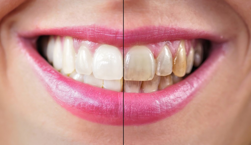
How To Get Your Smile Camera-Ready

Preparing for an event, beach party, or a photoshoot? There’s a lot to think about, right? There’s your outfit, the venue, transportation, food and drinks, and more. But don’t fret! You will have a great big day for sure. At the end of the day, you get to sit down, relax and look at photos taken by Boudoir Photography Edmonton. There’s plenty of opportunities to enjoy and show off your smile.
The question is – is your smile camera-ready? If your answer is anything else but yes, then you are in the right place! Here’s how you can get your smile shining bright for any special occasion.
Tip #1: Whiten Your Teeth
Whatever occasion or event you’re in, white is almost always a cornerstone of all color palettes around you. If you’re not confident about your smile, you may become self-conscious and ruin the day. That’s the last thing you want to happen. Gladly, a quick visit to your dentist may just help you with that. Many dental offices offer Teeth Whitening services for an instant and long-lasting photo-perfect smile.
If you don’t want or don’t have the budget for teeth whitening, you can always use at-home teeth whiteners including whitening toothpaste, mouthwash, strips, and gels. Including them into your dental hygiene regimen will protect and boost your smile for the camera.
Tip #2: Watch What You Eat & Drink
Are you a coffee lover like me? Unfortunately, this isn’t the best for our smiles. If you’re days or weeks away from an event, it’s better to stay away from foods and drinks that stain your teeth – coffee, wine, soft drinks, blueberries, and tea, among others. Don’t forget the biggest culprit of all – tobacco! I know it’s neither food nor drink but this is the holy grail so remember it.
What you can consume instead are foods rich in calcium (dairy products) and firm fruits and vegetables (celery and apples, for example). These all help keep your teeth bright and white for the occasion.
Tip #3: Brush Your Teeth Regularly
I know this is too simple of a tip, but regular tooth brushing is key to keeping healthy and clean teeth. Don’t brush too hard though as this could damage your tooth enamel and cause receding gums. To take away this human error, you can use an electric toothbrush that has rotating heads and maintains stable pressure to make sure each tooth receives the right care. Regular tooth brushing is sure to give you a bright smile.
Tip #4: Drink Plenty Of Water
Do you know that water has fluoride? Fluorine in water is added at a controlled and safe level for public consumption to help reduce tooth decay. This strengthens teeth, prevents cavities, and helps with overall oral health.
Tip #5: Wear Lipsticks The Right Way
You might wonder why commercial models in magazines use hot red lipsticks almost always. One of the reasons being, the contrast between the white teeth and the red lipstick makes the smile appear even brighter. But you can’t always wear red lipstick on all occasions, right? So, feel free to use other shades that compliment your smile best. Wearing the right shade and putting on a glossy finish can make your smile appear shinier and ready for the camera.
Final Thoughts
There’s a famous line that says, ‘You’re Never Fully Dressed Without A Smile’. So, whatever occasion you’re in, wear your best smile always! A perfect smile doesn’t happen overnight – but use these tips and you’re halfway to achieving it. Confidently show your white teeth with a natural and unforced smile to turn heads on any occasion. The camera will love you whichever angle it gets.
March 13, 2017
What Alcohol Really Does To Your Teeth?
Drinking alcohol may be enjoyable for many, but for some it can be a bit risky especially if consumed in large amounts and on a regular basis. Like drinking soda, alcohol contains certain enzymes that can damage the body over time. And in this particular case, alcohol can gradually damage the teeth.

And for a long time consumers of alcohol, this can become a major problem as this would not only do their teeth run a greater risk of being damaged by alcohol, but also the difficulty of trying to get them to move away from alcohol. And if you’re one of those two more concerned about the state of condition, then a quick alcohol detox will do the job just fine. Where can you go to detox alcohol from your body?
Alcohol Abuse Can Cause Damage to your Teeth
First of all, chromogens attach themselves to the enamel of the teeth that is already exposed to the acid present in alcohol. This results in the teeth being stained.
Second, is that alcohol can dry the mouth. Saliva keeps the mouth as well as the chief moist which removes the plaque. And without moisture, plaque and bacteria build up in the mouth which then digs into the teeth, causing damage.
As stated earlier, to detox alcohol from one’s body, it may take a considerable amount of knowledge and assistance. We can look around through the internet as much as you want, but you will find very little. But fortunately, Detox Matrix is just the one you need.
Safe and Effective Method
 Detox Matrix teaches new users the various methods and strategies to successfully detoxify alcohol from your body. This is especially useful for heavy drinkers, as they are most vulnerable to damage if left unchecked.
Detox Matrix provides many individuals who suffer from alcoholism the opportunity to cleanse their bodies, thus, giving them the first that they need in order to live a healthier lifestyle starting with their teeth.
November 2, 2016
Fluoride | The Perfect Coating For Your Roof?
Just like a dentist, a good workman uses only the best tools. So when it comes to roofing, you’ll want to utilize a coating that works effectively, is of high quality, and has excellent durability. Essentially, if you want to turn roofing leads into sales and deals and ultimately make a name for yourself, you’ll need products that can convince a wide range of buyers. That is where fluoride comes in handy in other professions. Well, more specifically, Polyvinylidene fluoride.
Roofing Salesman prides itself in using PVDF roof coating for its roofing. What exactly is this coating? Essentially, it’s a water- and fluoride-based coating that is reflective and repels dirt to high degrees. Its high quality makes it the perfect choice for Roofing Salesman, and it’s the reason that roofing leads can become clients.
So how does fluoride work so well in terms of roofing? Its benefits are exceptional and make it the perfect coating for any roof. Here are some of its winning characteristics.
1. Maintains reflectiveness
Most roof coatings collect dust and dirt, which accumulate over time and ultimately start to reduce the reflectance of the roof, which leads to the roof then becoming hotter and absorbing more heat. The weather also may corrode or diminish reflectance, and this can be troubling for anyone who wishes to make their roof fully durable. However, fluoride can repel all these factors, paying no heed to collected dirt, repelling unwanted particles, and withstanding all forms of weather and temperatures. It keeps the reflectance levels high regardless of the environment.
2. High resistance
 This sort of coating displays a high level or resistance towards standing water and mildew, and it ultimately lasts for a long time due to its tolerance of varying degrees of weather and temperatures. With that being said, as much as possible, try not to apply a fluoride-based coating in conditions that have ponding water – there’s no fixed guarantee that this will work, although this coating has been proven to be stronger and more resistant than most water-based coatings. In fact, Polyvinylidene fluoride is often found used in electrical wires insulation; this is because it is lightweight, has low thermal conductivity rates, is resistant to chemical corrosion, and is extremely flexible. That’s a selling point for sure, and it’s why it’s a top pick for roofing leads.
3. Compatibility
A fluoride-based coating, due to also being water-based, is compatible with a wide range of products. Water-based sealants and coating products can be used in collaboration with a fluoride roof coating, meaning it’s not necessary to search high and low or go out of your way to find product that works in tandem with it. When you purchase roofing leads, potential clients will be happy to see plenty of options available to them.
4. Environmentally friendly
Sure, this product might be fluoride-based, but it’s also water based. This means that it is more friendly to the environment than other coatings that are not. In addition, because it generally lasts longer, it won’t need to be touched up or redone often – some sellers even offer a 20 year guarantee. That’s how reliable and durable fluoride is when it comes to roofing! This adds excellent marketability to the product, making it easier to get roofing leads who stick around to make a deal.
5. Fast and easy
The nature of fluoride-based products make it easy to clean up in case of a mess, and it also cures quickly. To secure roofing leads and keep customers hooked, you’ll want to be able to guarantee that work can be finished as quickly as possible and without causing too much of a mess. A  PVDF coating dries and cures much faster in comparison to a lot of other water-based coatings, so it makes a perfect pick for this situation.
Need more proof of the effectiveness of Polyvinylidene fluoride in roofing? You don’t have to take my word for it. Famous buildings around the world make use of this form of coating, including renowned sites like Taipei 101 in Taiwan and the Petronas Twin Towers in Malaysia. With buildings that tall, coatings must work perfectly, and they clearly do. The proof is all around you. That’s why Roofing Salesman uses PVDF roof coating; you can’t deny that it works, and works well.
January 17, 2016
Fluoride In Weight Loss Pills? The Rumors Aren’t True

With the ever escalation of the need to lose weight, the Forskolin belly buster has come in handy to lend a helping hand in this process. It has been designed to help one lose weight while not compromising on their health. This is because some people have had to resolve to life-threatening ideas in an attempt to reduce that belly. This is the reason that prompted creation of Forskolin for weight loss, which ensures one is safe from all the risks associated with the other poor weight loss methods.
However, in the desperate attempt of Forskolin to curb the menace associated with increased weight, critics have come up with rumors that one of the ingredients is fluoride. These rumors have elicited mixed reactions and heated debates on the safety of these weight loss pills. However these rumors remain just that; rumors. This is because this product employs excellent scientific and technical know-how in its creation.
It is however worth noting that the adverse effects of fluoride are catastrophic. If present, it could have resulted to serious implications. For instance, use of fluoride has scientifically been found to cause adverse effects to the brain neurons. This could lead to serious brain infections and the consequences are too deadly for anyone to contemplate. With this kind of effects on the brain, there is no way any reputable company making products like the Forskolin belly buster would include such an ingredient in a product meant to help a person lose weight.
 Unfortunately there are some companies that do allow fluoride in their multivitamins, even though it is crystal clear that ingestion of fluoride results in slow bone formations. This could also interfere with the normal development of the bones. This is a very dangerous effect since it affects the overall well being of an individual. It also results in gastrointestinal discomforts. This in one way or the other leads to serious health implications. For that reason, ingestion of fluoride is catastrophic and should never be included in any ingredients meant to be ingested.
It is at this juncture that we need to look critically at the effects of this poisoning product; fluoride. Having stipulated all these effects, you can be sure that forskolin belly buster has not even a trace of fluoride. It is medically tested and there is no doubt that it has passed all the required tests and approvals.
December 30, 2015
Grand River Dental, Lacrosse
When I was born, I didn’t have any lateral incisors on the left or the right. It is for this reason that my canines were closer to the center than they are for most people. This abnormality was something which I could really do anything about since I was born with it. But it did make me look a lot like a vampire. This is the reason why I have hated my smile all through my life. Everyone told me to visit a dentist La Crosse Wi and get this problem fixed but I was too scared. I keep telling myself that there is nothing to fear and that everything will be alright. Unfortunately, I was still too worried even thinking of another person putting sharp instruments into my mouth. What made matters even worse was the fact that people kept telling me about how they felt dentists were dangerous and how I would need to suffer a lot if I were to fix this problem.
 It was only recently that I built up the courage to finally visit a dentist. This was after one of my co-workers recommended GRD Lacrosse. I took his advice and decided to call them up for an appointment. It was from this moment on that I knew I had made the right choice. They were just so professional in every aspect. If all dentists are like this, it’s hard for me to believe why anyone wouldn’t want to visit them.
Don’t think of this as a professional GRD Lacrosse review. That’s not what it is. This is just my honest opinion of their service and facilities.
What impressed me the most was the fact that the staff were extremely courteous. They weren’t in a hurry to move from one patient to the next and were quite pleasant. This is not what I was expecting. When I met the dentist who was going to look at my teeth as well, he spoke to me very nicely and took no time in making me feel comfortable around him. He assured me he could fix my teeth and explained the entire process to me in detail as well. And if that wasn’t enough, he answered every single question I had as well. What more could you want?
My permanent veneers were fitted into my mouth just 7 days later. When I smiled for the first time after the operation, I was so overcome with joy that words weren’t escaping my mouth. I thanked the dentist for their work and was more confident than ever. I am now going to spend my entire life telling people what a great job the GRD Lacrosse team did on my teeth.
Just visit the place and you will agree with everything I have said from the moment you enter the doors. You will see how the dentists there are all perfectionist. What are you waiting for? Don’t let dental problems hold you back any more. Just pick up the phone and set up an appointment with them today.
Address & Phone
Grand River Dental
305 3rd Street South
La Crosse, WI 54601
☎ 608-498-4660
F: 608-260-7706
December 2, 2015
Dentists Finding New Hobby That Anyone Can Participate In
If there were possibly one activity or social institution that could rival football, it would probably have to be Premiership fantasy football. The world of fantasy sports is huge all around the world both domestically as well as internationally. As more and more groups are starting to see their own communities form little enclaves within the football world, the reasons for dentists to join the fun has become to become deafeningly apparent. Dentists are starting to see that the fantasy world isn’t as far away from their world of health, healing and medicine.
Controlling the roster and lineup of a fantasy team is a challenge that certainly isn’t too simple for the finer minds of medical professionals. Dentists have the pick their squad and face off based on the real-world stats going down in each real world match as all other player do and there are some serious benefits for this on behalf of dentists.
The time is right for dentists to play
Everybody needs a hobby for but for dentists looking to get involved in premiership fantasy  football, the hobby takes on a bit more significance. For the dentist, there can be this “crystal castle effect” where the dentist feels isolated from some of the other people in the world due to the exclusion and loneliness that goes into learning a trade and a craft such as dentistry. Getting into a hobby or social group activity that breaks down this position of solitude is definitely a great thing.
Don’t forget, dentists have some serious downtime when there aren’t enough clients to help on a given day. Granted for non-dentists that might seem so unreasonable but for dentists, they can attest to the swings of work load on the job. Taking some of that downtime and translating it into time optimizing and
How will this look?
As a result, dentists go out of their way to find ways to bridge the gap between themselves and some of  the rest of the world. One thing they and nearly everybody shares in common is the love for sports and football is chief among those sports. Acting on this natural love for sports action and competition, people can find ways to share moments and common grounds, rather than continue to find ways to keep people apart. Despite the competition that is inherent in fantasy premiership leagues, there is also a real group camaraderie that exists between the various participants. This is not to mention the robust community of forums, websites, social media and newsgroups around the practice of playing fantasy premiership football. “Seek and ye shall find,” as it were.
Even on an individual level, dentists have a decent amount of discretionary spending potential  compared to the average person. Knowing this and having this true flexibility allows dentists to be able
to add the extra and more exciting element of gambling into the equation. Betting on the outcome of a particular match or league ranking is a special ability for dentists and other professionals that many premiership fantasy football league players might not enjoy.
Getting started
Search out league sites such as PremiereLeague.com and you’ll be able to find nearly any specific type of league you seek. Leagues with dentists are available as well as specific leagues that cater to pre-med dentists. Or, if you don’t see the league you want, many premiership fantasy football players will simply just create a league that has the features or culture they want. That’s the advised path to take so that other people can see the group you’ve created and possibly join it.
As dentists, you’ll find a way to be among your peers or in a mix of people from all walks of life in the online fantasy leagues, depending on which teams you are interested in and how much you want to wager. The world might have a certain perception about you as a dentist but often that is not really accurate. The truth in the matter is that while dentistry doesn’t have a specific outlet for competition, the world of online premiership fantasy football offers that outlet for dentists. I certainly don’t want my dentist to be trying to beat his own best time or compete against other dentists for “fastest root canal” honors. Much better is to have these excellent online spaces set aside for dentists to find some recreation on the side of a caree
November 11, 2015
Dentist Has Completely Uncanny “Tooth” Related Wedding.
So I’ve definitely heard of some unusual wedding entertainment ideas, but when I heard that two of my dentist friends from college were going to wed, I fetched my diary and was checking if I had time to attend the friends’ wedding when the informer added: They are going to have a completely tooth health-themed wedding. I closed the diary without checking my schedule and resolved to attend in spite of any appointments I was going to have that day. It wasn’t so much out of love as it was out of curiosity. Dentist-themed weddings are clearly unheard of, so I wanted to know what kind of craziness had come upon my friends.
The first bout of surprise was when I saw ribbons hung from the fibers of toothbrush imitations. The ribbons themselves looked like a spurt of toothpaste and were in different colors- yellow, white, orange, purple et cetera. Beside each ribbon hung a balloon. On each balloon was an immaculately done drawing of a couple with sparkly, well-aligned teeth, kissing. There was a burst of colors outside, a harbinger of the splendor and immaculate themes inside.
Similarly done balloons and ribbons hung from the ceiling of the chapel. Looking around, the chapel bore the aura of a dental clinic. On the aisle, the couple sat as dental patients would waiting for an appointment with a dentist.
 Across them, a giant tooth cake stood on a pedestal with a male-looking tooth beside a female-looking tooth, both showcasing radiant smiles. On top of the cake, where cake toppings should be, were the bride and groom as teeth. I would later be told that the cake didn’t have a lot of sugar as sugar spoils teeth.
The couple had asked a dentist who was also a church minister to preside over the occasion. He wore a dentist’ overalls that had the typical clergy’s collar tab. He looked amazing, albeit unusual, in that attire.
I sat there mesmerized by the creativity and passion of the couple. They had surpassed expectations already. I had come here cynical of a dentist-themed wedding, but now I was awed beyond expression. The couple looked fantastic in their wedding attire. The bridegroom and his grooms were dressed impeccably in well-done grey suits while the bride was gorgeously clad in a snow-white overflowing wedding dress. Her bridesmaids were just a gorgeous. Surprisingly, there was nothing dental in their various attires save for breast pocket handkerchief in men’s coats that hung from the pockets as would a tongue out of the mouth, though not as goofy, but in a stylish, decent way.
The height of this dentist couple’s ingenuity showed when the minister stood to have them take their vows. I cannot remember exactly what happened, except for a few interesting snippets, but I can firmly attest that it was one of the most spectacular things I have ever witnessed. It was unusual, genius and spectacular, to say the least. For instance, in the vows he asked the couple to stick together as teeth does to gum. When the teeth falls off, he said, it is to its own destruction. The minister asked them to seek counselling whenever they have marital problems just as people seek dental care whenever they have dental problems. When the couple partook of the cake, they brushed their teeth before the audience amid rapturous cheers.
As I drove off from the wedding, I looked at the diary: I was to meet the state’s governor the very time I was attending the wedding. I smiled to myself, thankful that I didn’t look at my diary in prior as I would have been compelled to go to the meeting with the Governor and miss the spectacular wedding.
November 8, 2015
5 Reasons Why Dentists Need To Create An App For Their Business
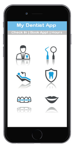 If you are a dentist, it’s highly recommended that you consider creating an app for your business. In this age wherein most people are connected to the internet, it makes complete sense that you reach out to your clients online. There’s a lot of benefits that you can reap from a simple app that’s available for both desktop and mobile users. With that said, now is the perfect time to develop an app for your dental clinic and separate yourself from your competitors. If you still have doubts about getting an app, the following reasons should help you understand why you should have one.
1) It will be a lot easier for clients to book for appointments.
If you have an app, a client can set up an appointment without visiting your clinic. It doesn’t matter if you and your clients are miles away from each other, business will continue as usual. Most apps these days are automated which means you don’t even need to move a finger to manage your appointments. Everything will be handled by the app. Apps are usually equipped with a scheduling system which enables clients to see what times the dentist will be available for appointments.
2) It’s a great way to build your brand and improve your reputation.
An app is very effective in getting the word out about your clinic and your dental services. In effect, your established clients will market and promote your business by simply using your app and recommending it to their friends and families. That’s like hitting three birds with a single stone. Additionally, people are known for having more trust in businesses that are active and visible online.
3) It’s very affordable.
Whether you are creating the app yourself or you are hiring someone to create it for you, the total cost won’t cut deep into your wallet. The investment you make is small compared to the benefits that you are going to reap once your app is launched. With the growing number of developers today, the cost of having an app created will likely go down in the coming months.
4) It’s a good way to attract more clients.
As we mentioned earlier, a lot of people these days are now using their laptops and mobile phones to search for products and services online. Needles to say, if you are not visible online, then you are not getting a slice of this growing online market. However, if you have an app, there’s a way for potential clients to find you and the dental services that you are offering.
5) It cultivates customer loyalty.
You can also use your app as a portal for customer service. Your current patients can use it to contact you about problems they may be having with their teeth, gums, or dentures. This connectivity builds loyalty among your customers. This means they are more likely to use your services again and even recommend you to people they know.
In conclusion, you need an app to market and promote your clinic and your dental services. It’s a good way to get ahead of your competitors.
November 6, 2015
A Hidden Danger In Swimming Pools
Fluorine is a naturally occurring element mostly found in water bodies for instance seas, lakes, rivers and even swimming pools. Fluorinated swimming pools in Arizona expose the swimmers into various health risks even though fluorine as a chemical is credited for reducing tooth decay thus many dentists recommends for its use in toothpastes. Pool Resurfacing Phoenix company offers a solution by eliminating all health risks posed by fluoride in Arizona swimming pools. Below are the top 10 health risks that are likely to originate from fluoridated swimming pools.
Top 10 health risks of fluorinated water
1. Reproductive problems
High levels of fluorine in the body reduce sperm count in males and ultimately leads to infertility. This is a proof that has been provided by scientists after research.
2. Brain damage
Fluorine is among the top 100 chemicals that are known to cause neurotoxicity to humans as depicted in the US Environmental protection Agency. Brain damage alters normal brain function.
3. Lowered IQ
When 1.9 parts per million accumulates in the body system, then chances of mind retardation is very high and ultimate lowered IQ happens.
4. Bioaccumulation of fluorine in the body
When ingested, fluorine is excreted through kidneys. But small quantities of fluorine tend to accumulated in the bones. This reduces the integrity of the bones in terms of strength. Pool resurfacing Phoenix helps to prevent such incidence from happening by cleaning fluoride elements from swimming pools.
5. Dental fluorosis in bottle fed children
Bottle fed children consume liquid foods. They thus have high chances of developing dental fluorosis due to fluorine contained in their meals.
6. Reduced thyroid function
Fluorine reduces the activity of thyroid hormone and increased used of it causes hypothyroidism causing a serious health effect. Swimming pool maintenance by Pool Resurfacing Phoenix can halt this health risk.
7. Arthritis condition
Fluorine creates bones and joint diseases which are similar to rheumatoid arthritis. This condition is associated with pain in the joints.
8. Bone damage
The calcium matrix in the bone structure is interfered with by fluorine making the bone weak. This problem is caused by high accumulation of fluorine in the bones.
9. Hastens osteoporosis amongst the elderly
Fluorine causes hip fracture in elderly women. Consumption of fluorinated foods by old women accelerates the rate of developing osteoporosis.
10. Early onset of puberty in teenagers
Accumulation of high levels of fluorine in pineal glands reduces production of melatonin which in turn leads to early onset of puberty.
How to avoid fluoride
The best step to avoid dangers of fluoride is to clean the swimming pools as well as carrying out maintenance tasks.
Pool Resurfacing Phoenix is equipped with swimming pool cleaning experts who chemically balances the pools and reduces the dangerous levels of flouride that is harmful to humans. The company also does maintenance work to ensure healthy pool water is guaranteed. Filtering of piping systems that feeds the pool is of importance in elimination of fluoride in the swimming pools.
October 27, 2015
Smile Matters: Dentist Services Review
 Have you been trying to hide that missing tooth which happened because of an accident? Or do you feel forced to cover your teeth because they are too yellow? And if you have multiple problems with your teeth, can they be all dealt with under one roof effectively?
These are problems which many people face everyday. So, what is the solution?
Smile Matters is an organization which offers solutions to such problems and claims much more. Let us now try to find out if it is really worth your while.
Friendly atmosphere:
Dentist’s office is perhaps the most detested place by most people. This is so, not only because of the severe pain associated with dental problems, but also because of the attitude of the doctors and other staff. Smile-Matters seems to have grasped the import of this well. They have made it their motto to make the patient feel comfortable and welcome in their office. This definitely helps you to relax and also minimizes your discomfort during any procedure.
Services:
This is by far the most important consideration when choosing your dentist. Smile-Matters offers a very wide range of services, all under one roof. Right from fillings and root canals to complicated cosmetic procedures, all are being provided in one place. So you do not need to worry about having to go to a different dentist for each of the problems that you may have. The place also seems to enjoy a good reputation among the patients, most of whom are quite happy with the quality of work and the work ethos. They are willing to go back again for any problem they may have in future. This is surely the biggest compliment that any doctor can ask for, and it is more true in the case of a dentist.
Equipment:
The importance of technology in today’s world cannot be over-emphasized. Therefore, it is important that the dentist’s office is well equipped with the latest machines in order to be able to tackle all kinds of dental problems easily and effectively. Smile-Matters appears to be keeping pace well, having such recent technologies like lasers, digital radiography among others.
Holistic approach:
Many doctors, while practicing their profession, tend to forget that they are dealing with real people and think of them only as their patients. This restricts the view of the doctor and prevents him from thinking about other regions of the body which could be responsible for the patient’s symptoms. Unless the doctor is open to such reasoning, the real cause and thereby an accurate diagnosis is often missed. Smile-Matters lays claim to a holistic approach to patients’ problems, and as much emphasis is laid on prevention as on treatment in the case of any individual patient.
In spite of best efforts, it is always possible that a patient may have certain problems. This can happen to anyone and at any place. However, the important thing is that a patient is properly heard, and a genuine attempt is made to sort the problem out quickly and to the patient’s satisfaction. Smile-Matters is one organization which does indeed endeavor to put this into practice.
Your smile is an important aspect of your persona. It helps you to befriend people and also elevates your own mood. But your smile is only as good as your teeth and therefore your teeth deserve the best possible care. Smile-Matters definitely seems well equipped for providing this.
Preet K. Sahota, D.D.S.
39572 Stevenson Place, Suite 131
Fremont, CA 94539
(510) 744-9009
October 27, 2015
Review of Dr H Peter Ku of Smile Matters

If you are from Fort Worth, then maybe dentist H. Peter Ku, D.D.S., PA may already be familiar to you.
This is simply because he is known to be the best dentist in Fort Worth. When I say the best, I really do mean the best. Dr. Ku as we call him has been attending to his patients for a very long time now that is more than fifteen years and the type of experience he has on this job is simply fascinating. On top of having the best knowledge to take care of your dental problems, Dr. Ku has embraced technology hence he uses the latest technology there is in dentistry to make sure that all his patients are satisfied with his work.
 Whatever your dental problem, Dr. Ku can make sure that you go back home with a different story. The type of service you get when you choose to visit Dr. Ku is special and all the patients are treated with respect and honor. Booking for a session with Dr. Ku has also been made available to all patients interested in his high quality services. All you need to do is make a call or visit his website through which you can easily send a message. When it comes to the cost of Dr. Ku’s high quality services, you need to know that you do not have to spend a fortune. Dr. Ku offers his services at pocket friendly prices.
Dr. Ku offers all sorts of dental care services. These include care such as dental implants and sedation dentistry. If you can take time to talk to some of the patients who have been served by Dr. Ku, you will realize that this doctor sure is the best. Families from all over the world have been served by Dr. Ku and the number of patients and families being served by this doctor is always rising. This can only mean that the type of service being offered by Dr. Ku is both of high quality and affordable.
What people say about you is very important especially if you are a professional and up to now, I have not heard anything negative about Dr. Ku. If you look at his website, you will realize that it is quality and very simple to navigate. This is one of the main reasons why people from all over the world have come to learn about Dr. Ku. All the information you need is made available on the website including the number to call. The “send message” command available on the website has been use by so many people around the world who have been facing various dental problems because they have been able to get online assistance.
Dentistry has come a very long way and technology has become a better part of this practice. A good dentist should be able to use the latest technology to make sure that his or her patients are properly served. Dr. Ku on the other hand knows the importance of technology in dentistry and how it has managed to improve dentistry services. When you walk inside his clinic, you will see all sorts of equipment that can be used to restore that wonderful smile. It is also important to note that Dr. Ku does more than just treat dental problems. This doctor also provide his patients with tips and advice on how to maintain healthy teeth.
There is more to dentistry than just plucking out rotten teeth. You need to understand that even little children face dental problems and according to how children are, the type of environment in which a dentist operates should be suitable. At Dr. Ku’s office, you will find an environment that is suitable for both the children and adults. This is one of the main reasons why so many families have trusted Dr. Ku for their dental problems.
There are so many dentists in this world but there are some dentists who you cannot compare to others and Dr. Ku is one of them. He believes in client’s satisfaction and he respects his profession. Take a look at his website and see what he has for you. Feel free to contact Dr. Ku as he is always waiting for you.
October 27, 2015
How To Determine Approximate Costs For Denture Work
 How do you determine what a reasonable cost for dentures would be? Like many orthodontic services, this depends on exactly what you need and where you are willing to go for these dentures. The typical cost of dentures is between $300 to $5000 per plate and many high-end denture models can cost between $1000 and $5000 depending on precisely what you’re looking for, so there is a bit of a trade-off between getting highly functional dentures that look like your natural teeth and finding a more “affordable” denture that costs $300 but may not be as durable or aesthetically pleasing. However, that doesn’t mean you need to overpay for denture options that you can’t afford and don’t really need if you can find a workable denture within your preferred price range.
That makes setting a reasonable budget worth the extra effort even though you might think that it’s “just” a denture. When you shop for dentures, you want to decide how much you are willing to spend when discussing options with your orthodontist so that you don’t get talked into something you won’t be able to afford. Ask for prices up front and the ones that are within your set budget are going to be your real options. While it may be worth it to spend an extra $100 for a more realistic looking denture or one with reduced maintenance needs, this eliminates the hidden sticker shock involved in shopping for dentures.
The amount of money that the orthodontist charges can be one major factor in the total cost of your dentures. You’d be surprised by how much this can vary between orthodontists in your area and those costs may even vary between region due to variances in the cost of doing business that orthodontists have to factor into the amount they charge. While you always want to makes sure you’re comparing apples to apples when looking around for an orthodontist and that includes asking for reference or looking for reputable review sites that feature orthodontists, it pays to get a few quotes from orthodontists in your area before you get dentures.
Most of us who have health insurance also have insurance that covers routine visits to the dentist, but is that insurance policy going to cover dentures? This is one common insurance “gotcha” for people who need dentures or any other kind of specialized orthodontic service. Making sure your dentures are covered before you go to the orthodontist can help to keep your out-of-pocket expenses down.
Dentures are not cheap, but shopping for orthodontic procedures like denture installation using many of the same tactics that you would use while shop for any other major purchases will help to keep the costs under control. That means knowing what you need in advance and what a reasonable price range would be regardless of whether you’re just looking to have a single tooth replaced by a denture or need a full bridge. It might also include visiting several orthodontists in your area to collect quotes and following up on the references they provide to see whether other real clients were happy with their work.
For more information please visit the American Dental Association.